Τρίτη 27 Σεπτεμβρίου 2011

Χονδροπάθεια Επιγονατίδος

Χονδροπάθεια Επιγονατίδος
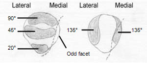
Η Ανατομική του Γόνατος
Η Χονδροπάθεια του Γόνατος  είναι μια συχνή επώδυνη πάθηση των γονάτων. Στην αμερικανική και αγγλική βιβλιογραφία, η πάθηση αυτή αναφέρεται με τον όρο Pattelofemoral Stress Syndrome το οποίο εμπεριέχει την Χονδροπάθεια Επιγονατίδος   την Χονδροπάθεια των Μηριαίων κονδύλων αλλά και τις Τενοντίτιδες του τετρακέφαλου και του Επιγονατιδικού συνδέσμου.
Παθολογική Ανατομική


H Χονδροπάθεια του Γόνατος προσβάλει κυρίως τον χόνδρο της επιγονατίδας και κατά δεύτερο λόγο το χόνδρο της Μηριαίας τροχιλίας. Ο χόνδρος των Κνημιαίων κονδύλων δεν είναι εύκολο να φθαρεί γιατί προστατεύεται από τους δύο Μηνίσκους, οι οποίοι όταν φθαρούν ή αφαιρεθούν οδηγούν σε φθορά του χόνδρου των Κνημιαίων κονδύλων.
Επιδημιολογία
Η Χονδροπαθεια της επιγονατιδας, είναι συχνή επώδυνη πάθηση του γόνατος η οποία εκδηλώνεται συνήθως σε νέα άτομα από 15-40 ετών. Προσβάλλει περισσότερο τις γυναίκες παρά τούς άνδρες, σε αναλογία 3:1. H Χονδροπάθεια είναι πιο συχνή από τούς τραυματισμούς των Μηνίσκων, αλλά συνήθως παραμένει ασυμπτωματική στην εφηβική ηλικία, διότι αυτοϊάται με σχηματισμό ινώδους χόνδρου. Η πάθηση είναι αρκετά συχνή ακόμα και σε άτομα εφηβικής ηλικίας. Υπολογίζεται ότι το 80-85% του πληθυσμού στην ηλικία 20-30 έχει εκφυλιστικές αλλοιώσεις και βλάβες του χόνδρου της επιγονατιδομηριαίας αρθρώσεως

Αιτιολογία

Η Χονδροπαθεια της επιγονατιδας, οφείλεται σε είτε σε τραυματισμό, είτε σε χρόνια υπερφόρτιση του χόνδρου της Επιγονατίδας, είτε σε εσωτερικούς παράγοντες γενετικής προδιάθεσης («ευπάθειας») του αρθρικού χόνδρου, είτε σε συνδυασμό αυτών. Γενικά, η πάθηση είναι πολυπαραγοντική, οφείλεται δηλαδή σε συνδυασμό πολλαπλών αιτίων, τά οποία χωρίζονται σε ενδογενή και εξωγενή .


Στάδια
H  Χονδροπάθεια του Γόνατος έχει τρία παθολογοανατομικά στάδια:
Στάδιο Ι: Οίδημα του Χόνδρου με κιτρινόφαιες μαλακές και προεξέχουσες εστίες χωρίς καμία ελαστικότητα και κάμψη.
Στάδιο ΙΙ: Ρωγμές και «ξεφτίσματα» του χόνδρου. Ελεύθερα χόνδρινα σωμάτια στο αρθρικό υγρό και αλλοιώσεις χρόνιας Υμενίτιδας.
Στάδιο ΙΙΙ: Ελκη κατά τόπους στον χόνδρο. Το υποχόνδριο οστό είναι ακάλυπτο στον πυθμένα τους και υπόσκληρο.
 Συμπτώματα και κλινική εικόνα
Συνήθως, μετά από κάποιο τραυματισμό της Επιγονατίδας, ή από χρόνια υπερκαταπόνηση σε εργασία σε γονατιστή θέση, ή σε γυμναστήριο, ή χορό, ή σε πεντάλ τροχοφόρων, πρέπει να περάσουν 3-6 μήνες ώσπου να εκδηλωθούν τά φαινόμενα της Χονδροπάθειας στον ασθενή. Ο ασθενής αναφέρει ότι «έχει ένα πόνο μπροστά στα γόνατα», ότι «αισθάνεται ένα κάψιμο», ότι «ακούει ήχους κράκ στο γόνατο», ότι «του φεύγει το γόνατο στο κατέβασμα σκαλοπατιών» (σημείο Giving way), ότι «θέλω να τεντώνω τα γόνατά μου», ότι «δυσκολεύομαι να κάνω βαθύ κάθισμα», ότι «δυσκολεύομαι όταν σηκώνομαι μετά από πολλή ώρα καθίσματος» («σημείο κινηματογράφου»), κλπ. Η Χονδροπάθεια επιγονατίδων διαγιγνώσκεται με το ιστορικό και με την κλινική εξέταση με τα χέρια. Το πλέον αξιόπιστο τέστ είναι η "δοκιμασία ολίσθησης και πλάγιας μετατόπισης της επιγονατίδας" πάνω στη μηριαία τροχιλία, με το γόνατο σε έκταση και χαλαρό, οπότε αναπαράγεται κριγμός και πόνος. Αλλα κλινικά ευρήματα είναι οι προκλητοί πόνοι στα χείλη της επιγονατίδος, η ύπαρξη υδράρθρου και υμενίτιδας, το "Τest Zohlen" (πόνος σε σύσπαση τετρακεφάλου) και το "σημείο αναπήδησης", που είναι το απότομο τίναγμα της επιγονατίδας σε κάμψη 30-40 μοιρών


 Απεικονιστικός έλεγχος


Η πάθηση δεν φαίνεται στις απλές ακτινογραφίες. Το μόνο που προσφέρουν είναι η απεικόνιση κατασκευαστικών ανωμαλιών όπως η υψηλή θέση της επιγονατίδος, η αβαθής Μηριαία Τροχιλία, η Διφυής Επιγονατίδα, η έξω θέση της Επιγονατίδας, η ύπαρξη κύστεων, τυχόν ύπαρξη οστεοφύτων, κλπ. Ενίοτε βοηθάει διαγνωστικά η CTκαι η MRI, αλλά δεν μπορεί πάντα να απεικονίσει τις αλλοιώσεις του χόνδρου. Η Αρθροσκοπηση είναι η άριστη μέθοδος για τεκμηρίωση της διάγνωσης, αλλά απαιτεί γενική νάρκωση και νοσηλεία μιάς ημέρας. Πάντως, με το καλό ιστορικό, την καλή κλινική εξέταση και τις ακτινογραφίες, η διάγνωση τίθεται με βεβαιότητα 65% στο 1ο στάδιο και 80% στο στάδιο ΙΙ, χωρίς άλλες εξετάσεις
             
Θεραπεία
Ελαφρές περιπτώσεις χονδροπάθειας επιγονατίδας συνήθως αυτοθεραπεύονται μέσα σε 30-45 μέρες. Τα περιστατικά ασθενών, αντιμετωπίζονται με αδυνάτισμα σε παχύσαρκους, ελαστική περιγονατίδα με οπή, ανάπαυση, αποφυγή καταπόνησης σε σκαλοπάτια ή έντονη άθληση, αντιφλεγμονώδη φάρμακα (για τον πόνο και το οίδημα), και ανακουφιστικές φυσικοθεραπείες. Το ποδήλατο με μεγάλη κάμψη γόνατος βλάπτει. Οι ασκήσεις τετρακέφαλου δεν πρέπει να ξεπερνούν τις 30 μοίρες, στην οξεία κρίση της παθήσεως  Μεσαίας βαρύτητας περιστατικά χρειάζονται 5 ενδαρθρικές ενέσεις Sodium Hyaluronate,. H χρήση του Υαλουρονικού Νατριου από το έτος 1997 στήν Χονδροπάθεια Επιγονατίδος απεδείχθη λίαν επιτυχής. Φαίνεται να αναστέλλει τις βλάβες αλλά και να διευκολύνει την επούλωση στις ήδη προϋπάρχουσες, με αποτέλεσμα τα τελευταία χρόνια να έχουν σχεδόν εξαφανισθεί οι εγχειρήσεις γιά θεραπεία Χονδροπάθειας.
Σοβαρές περιπτώσεις Χονδροπάθειας της Επιγονατίδας μάλλον δεν θεραπεύονται ποτέ 100%, αλλά αρκετά θεραπευτικά μέτρα είναι αποτελεσματικά για την ανακούφιση των ασθενών. Η θεραπεία χρειάζεται, όχι μόνο γιατί πονάει ο ασθενής, αλλά γιατί σταδιακά οδηγεί τα προδιατιθέμενα άτομα σε Οστεοαρθριτιδα του γόνατος. Τα άτομα αυτά χρειάζονται συνήθως Επεμβαση αρθροσκόπησης, με τρυπανισμούς για αναγέννηση χόνδρου, διατομή έξω καθεκτικού συνδέσμου, κλπ. Τα τελευταία χρόνια εφαρμόζεται πειραματικά και η αυτόλογη μεταμόσχευση καλλιεργημένων χονδροκυττάρων του ασθενούς μέσω διαδικασίας αρθροσκόπησης, όμως η μέθοδος απαιτεί τρείς εγχειρήσεις αρθροσκόπησης υπό γενική αναισθησία. Εκτος αυτου μπορούν να γίνουν εξειδικευμένες διορθωτικές χειρουργικές επεμβάσεις. Μερικές από αυτές είναι η "εγχείρηση Maquet" (ανύψωση Κνημιαίου Κυρτώματος), η εγχείρηση διατομής έξω καθεκτικού συνδέσμου, η μηχανική λείανση του χόνδρου, οι τρυπανισμοί, κάθετοι και πλάγιοι, η εκτομή της Υμενικής πτυχής, η οστεοτομία της επιγονατίδος,

ΦΥΣΙΚΟΘΕΡΑΠΕΥΤΙΚΗ ΠΡΟΣΕΓΓΙΣΗ

Η θεραπεία περιλαμβάνει τρία στάδια:
• Αντιμετώπιση της οξείας φάσης
• Αποκατάσταση
• Πρόληψη
Στην αντιμετώπιση της οξείας φάσης, όπως συμβαίνει σε όλα τα σύνδρομα υπέρχρησης, πρέπει ο ασθενής να διακόπτει όλες εκείνες τις δραστηριότητες που του προκαλούν πόνο. Σε αυτήν την φάση εφαρμόζουμε τεχνικές για την ελάττωση της φλεγμονής, όπως είναι η θεραπεία με Laser, ιοντοφόρεση με διάλυμα solu-medrol και xylocaine 2%, παλμικό μαγνητικό πεδίο (διεισδύει στο σώμα ομοιόμορφα, ανεπηρέαστο και επιδρά στην ενδοκυτταρική κίνηση των ιόντων, αυξάνει δηλαδή την διαπερατότητα της κυτταρικής μεμβράνης, με αποτέλεσμα την ελάττωση του οιδήματος και του πόνου, γρήγορη απομάκρυνση των προϊόντων του μεταβολισμού, αύξηση της παροχής του οξυγόνου στην περιοχή), ειδική επίδεση του γόνατος, ώστε η επιγονατίδα να συγκρατείται σε σωστή τροχιά κίνησης, ενώ σύμφωνα με το αποτέλεσμα του πελματογραφήματος ο ασθενής άμεσα πρέπει να φορά στα παπούτσια του ειδικά κατασκευασμένα πέλματα, ώστε να εξισορροπούνται οι δυνάμεις που ασκούνται στο επιγονατιδομηριαίο μηχανισμό.
Όταν υποχωρήσει η φλεγμονή και το οίδημα το επόμενο βήμα είναι η ισχυροποίηση των μυών της περιοχής, ιδιαίτερα του τετρακέφαλου μυός, που έχει αποδειχθεί ότι παίζει σημαντικό ρόλο στην αποκατάσταση αυτού του προβλήματος. Χρήσιμη είναι η εκπαίδευση με Biofeedback. Το πρόγραμμα συνδυάζεται με ασκήσεις διατάσεων ώστε να βελτιωθεί η ελαστικότητα και η λειτουργικότητα των μαλακών μορίων της περιοχής. Τέλος το πρόγραμμα ολοκληρώνεται με ασκήσεις ιδιοδεκτικότητας.
Σημαντικό μέρος της θεραπείας είναι η ενημέρωση και η επανεκπαίδευση του ασθενή ώστε να προλαμβάνει τυχόν νέα υποτροπή του προβλήματος στο μέλλον. Περιλαμβάνει επανεκπαίδευση στην σωστή στάση, συχνά διαλείμματα κατά την διάρκεια δραστηριοτήτων που επιβαρύνουν τις αρθρώσεις των γονάτων, πρόγραμμα ασκήσεων, κολύμπι, ενώ καλό είναι να αποφεύγεται η άσκηση με ποδήλατο.

Παράλληλα με την ανάπαυση και τη φαρμακευτική αγωγή (αναλγητικά, αντιφλεγμονώδη) του ορθοπαιδικού, κρίνεται επιτακτική η ανάγκη για φυσικοθεραπευτική αντιμετώπιση που επικεντρώνεται στα παρακάτω:
• Φυσικά μέσα (tens,E.M.S., laser) για την ανακούφιση των συμπτωμάτων και τη μυϊκή ενδυνάμωση (έσω πλατύ).
• Κινητοποίηση επιγονατίδας (ολισθήσεις) και γόνατος στα όρια του πόνου.
• Μάλαξη γειτονικών μυϊκών ομάδων.
• Ενδυνάμωση τετρακεφάλου με έμφαση στον έσω πλατύ, για την ευθυγράμμιση της επιγονατίδας. Ισομετρικές και ισοτονικές ασκήσεις σε κλειστή-ανοιχτή αλυσίδα σε σετ των 10 επαναλήψεων.
• Ασκήσεις ιδιοδεκτικότητας γόνατος.
• Κρυοθεραπεία. Μπορούμε να κάνουμε παγομάλαξη ή να τοποθετούμε ψυχρά επιθέματα τυλιγμένα με πετσέτα για 10 λεπτά. Φροντίστε να επαναλαμβάνετε τη διαδικασία έως και 8 φορές μέσα στη μέρα.
• Τεχνικές περίδεσης της περιοχής με ελαστικό επίδεσμο ή άλλα είδη taping.
Η κολύμβηση είναι πολύ καλή αθλητική δραστηριότητα για άτομα με χονδροπάθεια επιγονατίδας, λόγω των μειωμένων φορτίσεων που δέχεται η άρθρωση του γόνατος. Αντιθέτως δραστηριότητες που προκαλούν πόνο όπως η άνοδος-κάθοδος σκάλας θα πρέπει να αποφεύγονται.


Cervical spondylosis


Cervical spondylosis


Cervical spondylosis is a chronic degenerative condition of the cervical spine that affects the vertebral bodies and intervertebral disks of the neck (in the form of, for example, disk herniation and spur formation), as well as the contents of the spinal canal (nerve roots and/or spinal cord). Some authors also include the degenerative changes in the facet joints, longitudinal ligaments, and ligamentum flavum.


Spondylosis progresses with age and often develops at multiple interspaces. Chronic cervical degeneration is the most common cause of progressive spinal cord and nerve root compression. Spondylotic changes can result in stenosis of the spinal canal, lateral recess, and foramina. Spinal canal stenosis can lead to myelopathy, whereas the latter 2 can cause radiculopathy.


Rehabilitation Program


Physical Therapy


Immobilization of the cervical spine is the mainstay of conservative treatment for patients with cervical spondylosis. Immobilization limits the motion of the neck, thereby reducing nerve irritation. Soft cervical collars are recommended for daytime use only, but they are unable to appreciably limit the motion of the cervical spine. More rigid orthoses (eg, Philadelphia collar, Minerva body jacket) can significantly immobilize the cervical spine (see Special Concerns). The patient's tolerance and compliance are considerations when any of the braces are used. A program of isometric cervical exercises may help to limit the loss of muscle tone that results from the use of more restrictive orthoses. Molded cervical pillows can better align the spine during sleep and provide symptomatic relief for some patients.


Mechanical traction is a widely used technique. This form of treatment may be useful because it promotes immobilization of the cervical region and widens the foraminal openings. However, traction in the treatment of cervical pain was not better than placebo in 2 randomized groups.


The use of cervical exercises has been advocated in patients with cervical spondylosis. Isometric exercises are often beneficial to maintain the strength of the neck muscles. Neck and upper back stretching exercises, as well as light aerobic activities, also are recommended. The exercise programs are best initiated and monitored by a physical therapist.


Passive modalities generally involve the application of heat to the tissues in the cervical region, either by means of superficial devices (eg, moist-heat packs) or mechanisms for deep-heat transfer (eg, ultrasound, diathermy).


Manual therapy, such as massage, mobilization, and manipulation, may provide further relief for patients with cervical spondylosis. Mobilization is performed by a physical therapist and is characterized by the application of gentle pressure within or at the limits of normal motion, with the goal of increasing the ROM. Manual traction may be better tolerated than mechanical traction in some patients. Manipulation is characterized by a high-velocity thrust, which is often delivered at or near the limit of the ROM. The intention is to increase articular mobility or to realign the spine. Contraindications to manipulative therapy include myelopathy, severe degenerative changes, fracture or dislocation, infection, malignancy, ligamentous instability, and vertebrobasilar insufficiency.


Σάββατο 24 Σεπτεμβρίου 2011

T4 Syndrome

T4 Syndrome

The thoracic spine region is comprised of 12 vertebrae and the rib cage with its articulations which enhance the stability of the thoracic spine (1, 2). In the vertebral column, the compressive load at T1 is about 9% of body weight, increasing to 33% at T8 and 47% at T12 (1). Although, there are many potential sources for pain at the thoracic region, research for this region remains limited (1, 2, 3). Various sources of thoracic pain that are musculoskeletal in origin can include: “muscle strain, vertebral or rib fracture, zygapophyseal joint arthropathy, active trigger points, spinal stenosis, costovertebral and costotransverse joint dysfunction, ankylosing spondylitis, diffuse idiopathic skeletal hyperostosis, intervertebral disk herniation, intercostal neuralgia and T4 syndrome (3)”. In this article, we will discuss T4 syndrome in more detail.

T4 Syndrome Symptoms
Symptoms associated with T4 syndrome include constant or intermittent upper thoracic pain which may also be accompanied with back stiffness and upper extremity numbness and/or paresthesia (3, 4, 5, 6). This paresthesia may be glove like in distribution for one or both forearms/hands (3, 4, 5, 6). Referred pain into the neck and scapular regions along with generalized headaches which are dull and achy may be associated with T4 syndrome as well (3, 4, 5, 6).

T4 Syndrome Description/Cause

The cause of T4 syndrome is inconclusive but may be associated with joint hypomobility and faulty postural alignment (2, 4, 5 ,6). Both the thoracic intervertebral disks and thoracic zygapophyseal joints are thought to be primary pain generators in T4 syndrome based on their pain patterns (5, 6). It is also hypothesized that sympathetic dysfunction somehow related to vertebral dysfunction in the upper thoracic region (T2-7) causes a referred or reflex phenomenon in the arm or hands (4). Women in the age range of 30 to 50 years of age are also 4 times more likely than men to develop this condition (3, 4, 6). Activities such as lifting, excessive bending, pulling, reaching, shoveling, performing overhead activities, prolonged sitting and poor posture can exacerbate this condition (4, 6).

Physical Therapy Findings in Patients’ with T4 Syndrome
Upon palpation and joint play assessment, patients will often display tenderness and hypomobility at and around the T2-T7 segments with the T4 segment being most involved (4, 6). A thorough neurological examination should also be conducted to rule out any neural involvement as the neurological examination presents normal in patients with T4 syndrome (4, 6).

T4 Syndrome Treatment Options for a PT
• Postural/Functional Training
• ROM exercises
• Stretching
• Strengthening/Stabilization
• Manual Therapy/Joint Mobilization
• Modalities (heat, ultrasound)

The Use of Continuous Passive Motion After Total Knee Arthroplasty

A continuous passive motion (CPM) machine is generally utilized as soon as possible postoperatively for aiding recovery after a total knee arthroplasty (TKA). The function of the CPM machine is to move the knee joint slowly through a controlled range of motion (ROM) to prevent joint stiffness and the development of scar tissue. It has been estimated by the president of an American company that makes and sells CPM devices that, as of 2002, CPM was being used in 17,000 hospitals in 77 countries and that about 7 million patients had been treated using CPM (1). Although the CPM machine may be a commonly prescribed treatment method, the effectiveness of its use remains conflicting.

In a review by Brosseau et al. (2), they conducted a meta-analysis of 14 studies to examine the effectiveness of CPM following total knee arthroplasty. Their study concluded that CPM combined with conventional physical therapy increased the active knee flexion of patients who had underwent a TKA by only 4° (and only at 2 weeks of follow up) compared to physical therapy alone. They also found through their meta-analysis that the use of the CPM machine decreased the length of stay for patients by 0.69 days and reduced the need for manipulation following TKA. In the face of these findings the authors did question the clinical significance of an additional 4° of knee flexion and cautioned on the interpretation of the length of stay data as many of the studies are historic in the terms of contemporary lengths of stay following TKA (3).

In a study by McInnes et al. (4) they evaluated the efficacy of CPM in postoperative management of patients undergoing TKA by conducting a randomized controlled single blind trial of 93 patients who participated in CPM plus standardized rehabilitation vs. standardized rehabilitation alone. Their study found that CPM patients achieved earlier motion and had 7° more active flexion on post op day 7 than those patients who didn’t receive CPM. However, by 6 weeks post op, the range of motion in the two groups were virtually identical.

In another study by Denis et al. (5), they conducted a randomized clinical trial to compare the effectiveness of 3 in-hospital rehabilitation programs for ROM in knee flexion & extension, functional ability and length of stay after undergoing a TKA. Eighty-two subjects were randomly assigned to 1 of 3 groups immediately after a TKA: a control group who received conventional physical therapy only; a second group which received conventional physical therapy and 35 minutes of CPM applications daily; and a third group which received conventional physical therapy and 2 hours of CPM applications daily. The results of their study revealed that adding CPM applications to conventional physical therapy interventions after TKA surgery didn’t favor increase knee ROM, functional ability or length of stay.

From a review of these studies we can conclude that no one study has been able to clearly demonstrate any long term benefit from the use of the CPM machine after TKA surgery. Besides the psychological benefits for a patient by using a CPM machine, conventional physical therapy services along with a home exercise program should be adequate for preventing joint stiffness and the development of scar tissue in most cases.

A Review of Patellar Taping for Patellofemoral Pain Syndrome

The patellofemoral joint is influenced significantly by the quadriceps muscle (line of pull, strength), shape of the trochlear sulcus, shape of the patella, surrounding soft tissue, biomechanics at the hip and foot, position of the tibiofemoral joint (quad insertion), pelvic obliquities/SI joint dysfunction, leg length differences, lower extremity flexibility and/or muscle imbalances (1).

“Compressive forces begin at 20-30 degrees of flexion between the medial and lateral facets of the patella and the femoral condyles. As flexion continues, the patellar articular surfaces segmentally come in contact with the trochlear groove with the exception of the odd facet. Continuation of flexion allows a smaller portion of the medial aspect of the patella to be contacted, with greater resultant compressive force. With extreme flexion, the odd facet comes into contact with the inner margin of the femoral condyle in the region of the intercondylar groove (1). Below is an image displaying the area where compressive forces are focused at the knee joint when performing knee flexion”.

It is generally believed that the alteration of patellofemoral kinematics can contribute to patellofemoral pain syndrome. Individuals with patellofemoral pain generally will have one of four patellar malalignments. Please see article Understanding Patellofemoral Dysfunction” by Kate Grace, PT, OPA-C & Annie Fonte, MBA” for additional information.

Interventions for treating patellofemoral pain syndrome (PFPS) can include patellar taping, patellar bracing, selective strengthening of the vastus medialis muscle, stretching tight lateral structures, correcting structural foot abnormalities, or a combination of these interventions (2, 3, 4, 5). In this article we will review patellar taping for the treatment of patellofemoral pain syndrome.

Patellar taping is sometimes utilized by physical therapists in conjunction with other treatment interventions when treating patients with PFPS. It is theorized that that most patients with PFPS would benefit from medialization of the patella which would off load the compressive forces at the lateral patellofemoral joint (2, 5). In a study by McConnell (5) she had a success rate of over 90% when incorporating taping to correct for a glide, tilt and rotational components of the patella along with emphasizing closed-chain exercises. A study by Derasari et al (2) utilizing dynamic magnetic resonance imaging revealed that taping resulted in a significant patellofemoral inferior shift which may account for some of the reported pain relief with taping, due to an increase in contact area. In another study by Gilleard et al (4), they found that taping the patellofemoral joint changes the onset of VMO and VL activity. The earlier activation of the VMO may promote VMO activity during retraining, improving patellar tracking.

Despite the positive findings in the above studies, other studies concluded that although taping the patella medially can contribute positively to PFPS rehabilitation, there were no significant changes in patella positions (6, 7). A study by Gigante et al (6) utilizing computed tomography found that patellar taping didn’t affect patellofemoral lateralization or tilt, while another study by Bockrath et al (3) utilizing Merchant’s view x-rays found no significant changes occurring with patella rotation, patellofemoral congruency, or sulcus angles. Although these studies didn’t reveal significant changes in patella position with taping, their subjects did demonstrate improvement with reduction of pain levels during physical therapy related activities. These authors hypothesized that the success of pain reduction from taping could be a result of increase sensory input from the tape, enhanced support of the patellofemoral ligaments and/or pain modulation via cutaneous stimulation (3, 6, 7).

Although studies may vary as far as taping the patella to correct for patella deviation, the majority of studies do concur that patients did experience some sympotomatic relief with taping and were able to better participate in physical therapy in order to allow for more aggressive functional rehabilitation and to facilitate compliance (2, 3, 4, 5, 6, 7).
Patella Taping Instructions (1)
1) Shave the area prn
2) Clean the area with alcohol prep
3) Position the patient in sitting with the knee in extension and the quadriceps relaxed
4) Apply white pre-wrap (Hypafix). This extends from the lateral epicondyle over the patella medially and posteriorly to the mid-popliteal fossa.
5) Apply the brown rigid tape as indicated below (8)
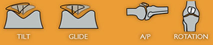
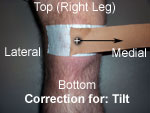
Lateral tilt component is corrected by pulling the tape from the midline of the patella medially.
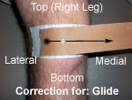
Excessive lateral tracking is corrected by pulling the patella medially.
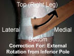
Excessive external rotation of the patella is corrected by taping from the middle inferior patellar pole upward and medially.
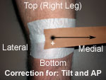
An anteriorposterior tilt of the patella is corrected by attempting to tilt the inferior pole of the patella anteriorly by placing a strip of tape at the superior pole of the patella and pulling the tape medially.

6) Symptoms should decrease immediately upon application. If present, always correct for AP (+) tilt first, then the worst component next (1).
7) For tape removal, be sure to protect the skin. Peel of slowly and hold the skin down. Tape remover may be necessary. Be especially careful with patients who are elderly and/or who have frail skin.

Osteoartrits and physical Therapy


According to the Arthritis Foundation , osteoarthritis is the most prevalent form of arthritis in the United States. Nearly one-third of all American adults have X-ray evidence of osteoarthritis in at least one area of their body.
With osteoarthritis an individual’s joint is affected. Anatomically, a joint is formed when two or more bones connect and are held together by ligaments. Certain joints in our body are called synovial joints. The bones that form synovial joints are covered with cartilage. The joint is also surrounded by a tough fibrous tissue called a joint capsule. The joint cavity within the capsule is lined with an inner membrane called the synovial membrane. This membrane secretes synovial fluid which acts as lubrication for the joint. The image provided displays the multiple joints and capsules of the hands.
The cartilage of synovial joints serves two important functions; it acts as a shock absorber and provides a smooth surface for the weight-bearing bones. This cartilage provides a soft, flexible foundation and absorbs the forces of energy produced by joint movement.
In the human body, the hips, knees, shoulders, elbows, wrists, and thumbs are examples of synovial joints. These joints have more mobility than other joints, because of their unique structure. When the mechanical stress placed on the joint is too great, or the joint’s ability to withstand the stress is compromised, physical changes can occur in the cartilage of the joint.
Osteoarthritis, also known as OA, is a complex metabolic disorder of the cartilage and bones of certain joints in the body. In OA, cartilage begins to weaken, becomes frayed, and eventually breaks down. This breakdown of cartilage exposes the underlying bone resulting in the bones of the joint rubbing together. This exposure can lead to pain, the fragmentation of bits of bone and cartilage which breaks off and floats inside the joint, the development of bone spurs on the edges of joints, and the enlargement and inflammation of the bones themselves. The image provided displays an x-ray revealing osteoarthritis of the knee.
The exact cause of OA is unknown. However, there are certain risk factors that appear to be linked to a higher occurrence of OA. These risk factors include: family genetics, repeated trauma to the joint, athletes and people who have extremely physically active jobs, persons with certain bone disorders, and obesity. Excess body weight places additional stresses to the joints and may have adverse effects on the cartilage and shock absorption function of the joint.
Individuals with OA often complain of pain that they describe as a deep ache which often occurs even while resting. With movement, these individuals may experience crepitus, popping or grinding noises, within the affected joint. Joint stiffness and inflammation may also be experienced, and often increases with changes in the weather/humidity.
There are many treatment options available for osteoarthritis. The goals of treatment are to: reduce pain, increase mobility and weight-bearing tolerance, and to minimize any potential permanent disability. Physical therapy is a common treatment option for OA. A physical therapist will assist in developing exercises that are safe and healthy for your joints and body. These exercises can be an excellent way to help keep joints mobile, decrease pain, increase body strength, and decrease overall body weight. Click here to see example exercises: Rehabilitative Exercises for Patients With Knee Osteoarthritis . Aquatic therapy and water aerobics is highly effective with people with OA as well. Aquatic therapy helps to decrease pain because of the therapeutic affect of warm water. Secondary to the buoyancy of the water, it also decreases the weight-bearing stress to the joint, therefore allowing one to move with less pain. If you are able to move through the water, your muscles can become stronger and be able to support your joint better. In addition, the resistance of the water provides excellent strengthening and toning of all of your body’s muscles, which can assist in overall weight loss. Click here to learn more about aquatic therapy exercises: Depth-Specific Aquatic Therapy: Why Pool Depth Matters .
In physical therapy, other treatments may include: paraffin (warm wax applied to the hands or feet), ultrasound, electrical stimulation, soft tissue mobilization/therapeutic massage, and custom foot orthotics. A physical therapist may also help a person with OA improve his/her mobility and walking by analyzing the movement and gait patterns while the person moves. This specialized analysis can help to decrease excess stress to a joint and improve overall mobility throughout the body


. ΚΙΝΗΤΙΚΟΤΗΤΑ ΤΩΝ ΑΚΡΩΝ – ΕΞΕΤΑΣΗ ΠΕΡΙΦΕΡΙΚΩΝ ΝΕΥΡΩΝ

(κίνηση, μυς, περιφερικό νεύρο, νευρική ρίζα)

· Απαγωγή ώμου 15 – 90 μοίρες: δελτοειδής, μασχαλιαίο νεύρο, Α5, Α6

· Κάμψη αγκώνα (σε υπτιασμό το αντιβράχιο): δικέφαλος, μυοδερματικό νεύρο, Α5, Α6

· Έκταση αγκώνα με αντίσταση: τρικέφαλος, κερκιδικό νεύρο, Α6, Α7, Α8

· Έκταση καρπού (πηχεοκαρπικής) στην ωλένια πλευρά: ωλένιος εκτείνων τον καρπό, κερκιδικό νεύρο, Α7, Α8.

· Έκταση καρπού στην κερκιδική πλευρά με τα δάκτυλα σε έκταση: μακρός κερκιδικός εκτείνων τον καρπό, κερκιδικό νεύρο, Α5, Α6.

· Έκταση δακτύλων στις μετακαρπιοφαλαγγικές αρθρώσεις: εκτείνοντες δακτύλων, κερκιδικό νεύρο, Α7, Α8.

· Kάμψη καρπού με κερκιδική απόκλιση: κερκιδικός καμπτήρας καρπού, μέσο νεύρο, Α6, Α7

· Κάμψη καρπού με ωλένια απόκλιση, ωλένιος καμπτήρας καρπού, ωλένιο νεύρο, Α7, Α8, Θ1

· Αντίσταση στην έκταση της άπω φαλαγγοφαλαγγικής του δείκτη: εν τω βάθει καμπτήρες δακτύλων, Α7, Α8

· Προσαγωγή/ απαγωγή δακτύλων χεριού: ραχιαίοι και παλαμιαίοι μεσόστεοι, ωλένιο νεύρο, Α8, Θ1

· Κάμψη ισχίου με αντίσταση (το γόνατο σε κάμψη και ο/η ασθενής πρηνής): λαγονοψοίτης, μηριαίο νεύρο, Ο1, Ο2, Ο3

· Έκταση ισχίου (ο/η ασθενής πρηνής): μείζον γλουτιαίος, κάτω γλουτιαίο νεύρο, Ο5, Ι1, Ι2

· Απαγωγή και έσω στροφή ισχίου (με το ισχίο και γόνατο σε κάμψη): μέσος και μικρός γλουτιαίος επιπολής γλουτιαίο νεύρο, Ο4, Ο5, Ι1

· Προσαγωγή ισχίου με αντίσταση: θυροειδές νεύρο, Ο2, Ο3, Ο4

· Έκταση γόνατος με αντίσταση (με το γόνατο σε κάμψη): τετρακέφαλος μηριαίος, μηριαίο νεύρο, Ο2, Ο3, Ο4

· Κάμψη γόνατος με αντίσταση: δικέφαλος μηριαίος, ημι-υμενώδης, ημιτενοντώδης, ισχιακό νεύρο, Ο5, Ι1, Ι2

· Έκταση μεγάλου δακτύλου με αντίσταση: μακρός εκτείνων το μεγάλο δάκτυλο, κοινό περονιαίο νεύρο, Ο5, Ι1

· Έκταση (ραχιαία κάμψη) ΠΔΚ (ποδοκνημικής): πρόσθιος κνημιαίος, κοινό περονιαίο νεύρο, Ο4, 05

· Έκταση δακτύλων με αντίσταση: μακρός εκτείνων τα δάκτυλα, κοινό περονιαίο νεύρο, Ο5, Ι1

· Πελματιαία κάμψη ΠΔΚ (ποδοκνημικής) (ή να σταθεί όρθιος/α στα δάκτυλα): γαστροκνήμιος & υποκνημίδιος, οπίσθιο κνημιαίο νεύρο, Ι1, Ο2

· Κάμψη τελικών φαλάγγων δακτύλων ποδιού: μακρός καμπτήρας των δακτύλων, οπίσθιο κνημιαίο νεύρο, Ο5, Ι1, Ι2.

3. Εξέταση ράχης (log roll στο τραύμα)

4. Εξέταση στα εν τω βάθει τενόντια αντανακλαστικά

5. Μυϊκή ισχύς:

0: παράλυση

1: ψηλαφητή ή ορατή σύσπαση

2: πλήρες εύρος κίνησης χωρίς βαρύτητα (οριζόντιο επίπεδο)

3: πλήρες εύρος κίνησης εναντίον της βαρύτητας (μειομετρική)

4: πλήρες εύρος κίνησης, αλλά λιγότερη από το φυσιολογικό

5: κ.φ. (φυσιολογική) μυϊκή ισχύς

Τετάρτη 21 Σεπτεμβρίου 2011


Σύλλογοι


Ιστοσελίδες Φυσιοθεραπευτικού Ενδιαφέροντος


KNEEguru (Θέματα και Πληροφορίες για το Γόνατο)

Ίδρυμα Αρθρίτιδας (Θέματα και Πληροφορίες για την Αρθρίτιδα)

Spine Universe (Θέματα και Πληροφορίες για Παθήσεις Αυχένα - Μέσης)


Επιστημονικά Περιοδικά

Physical Therapy Journal (Publication of the APTA)


Σύλλογοι


Ιστοσελίδες Φυσιοθεραπευτικού Ενδιαφέροντος